
A vascular surgery team from the University of Southern California (USC) in Los Angeles demonstrated the feasibility of multidisciplinary direct revascularization of segmental arteries to prevent spinal cord ischemia (SCI) using novel endovascular or extra-anatomic bypass techniques in high-risk patients with complex thoracoabdominal aortic aneurysms (TAAAs) following a retrospective review of fenestrated or branched endovascular aneurysm repairs (F/BEVARs) over a five-year period, attendees of the recent Western Vascular Society (WVS) annual meeting (Sept. 9–12) were told.
The early work, which involves 12 patients undergoing either an endovascular or open approach, was hailed by audience member Benjamin W. Starnes, MD, chief of vascular surgery at University of Washington Medicine in Seattle, who said, “This is a new idea. I applaud the courageousness of our colleagues at USC for pursuing this.”
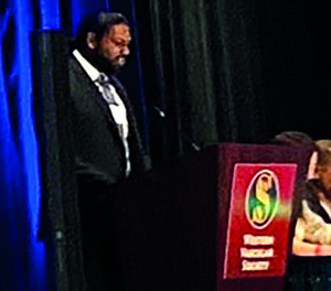
USC integrated vascular surgery resident Anand Ganapathy, MD, who presented the study results, told WVS 2023, held in Koloa, Hawaii, that nine patients were treated endovascularly, with a total of 11 segmental arteries incorporated. Ganapathy outlined how seven were revascularized using a directional cuff sewn directly onto a physician-modified graft that was extended with self-expanding Viabahn stents; two of the arteries were treated using stented fenestrations; and the remaining two—located along the seal zone—were tackled using unbridged, large fenestrations without covered branched stents. The other three patients received an extra-anatomic bypass.
Extents I and II were the most common type of TAAA among the patient group, Ganapathy continued. Procedural details for endovascular repair of the aneurysms demonstrated an average of 4.3 target vessels (2–6) treated, with a technical success rate of 83%. Failures (two patients) involved a bridging stent to a renal artery “that was dislocated from the fenestration and not recoverable” and a malalignment of an unstented fenestration to an intercostal artery “maintained with an uncovered stent.” Nine patients had successful prophylactic spinal drain placement. The three patients who underwent open repair of their segmental arteries received their bypass an average of three days prior to the F/BEVAR procedure.
Ganapathy reported no mortality at 30 days post-procedure. Two patients—one who had a staged extra-anatomic bypass, the other an unstented fenestration—incurred spinal cord ischemia that “resolved completely by discharge.” Both had prophylactic spinal drain placement. Average follow-up was 472 days. Three of the 14 targeted segmental arteries occluded during follow-up, Ganapathy added. Occluded branches involved one extra-anatomic bypass and two directional cuffs. “They occurred beyond one year without causing spinal cord ischemia,” he said.
Ganapathy concluded: “Spinal cord ischemia remains a serious complication of F/BEVAR, despite current mitigation strategies. Our initial experience demonstrates feasibility of endovascular and extra-anatomic bypass for segmental revascularization in select patients. Further work is planned to improve patient selection through anatomic and dynamic assessment of spinal cord perfusion patterns, as well as the safety and efficacy of segmental artery revascularization procedures through a multidisciplinary, collaborative effort across expertise from a growing list of specialists. Our vision is that someday this might be serve as another adjunct to the current spinal cord ischemia mitigation protocols.”
Karthikeshwar Kasirajan, MD, a clinical professor of vascular surgery at Stanford University, who was the designated discussant of the paper at WVS, summarized the study’s headline findings, outlining how the novel techniques had been employed in 3.8% of F/BEVAR patients reviewed during the study period. Some 11% of the endovascular group and 33% of the open group developed transient paraplegia, he said. Kasirajan commented: “In my opinion, it should be noted that stent grafts have a lower instance of paraplegia when compared to open repairs in general. This is despite the fact that length of coverage is often more extensive as to require landing zones that are much more extensive than open repairs. Additionally, during open repairs we can reimplant intercostals. This cannot be done during stent grafts [implantation], hence intercostal revascularization may not be the holy grail in the prevention of paraplegia.”
Alan Lumsden, MD, chair of the Department of Cardiovascular Surgery at Houston Methodist in Houston, Texas, congratulated the USC team on the ground-breaking nature of their work, asking Ganapathy and colleagues to drill down on any guidelines they follow to determine which patients should receive the novel approach.
Senior author of the study, Sukgu Han, MD, co-director of the Comprehensive Aortic Center at USC’s Keck Hospital, said: “Patient selection is everything. There’s probably more value from putting this protocol together with people from different specialties, maybe actually in the characterization of the spinal perfusion and individual variability. Because, so far, really the only surrogate marker we are looking at, in really very few select patients, is the size and assumption that that carries dominant collateral to the spinal cord.”


