Brought to you by:


Moving limb preservation forward with Shockwave Javelin Peripheral Intravascular Lithotripsy

This advertorial is sponsored by Shockwave Medical.
Chronic limb-threatening ischemia (CLTI) represents the most advanced stage of peripheral artery disease (PAD) and carries a significant risk of major amputation if left untreated. Calcified tibial lesions remain among the most technically challenging targets for endovascular intervention, as conventional angioplasty balloons frequently fail to track through or adequately dilate these rigid segments.
Intravascular lithotripsy (IVL) has emerged as a safe and effective tool¹̛ ² for modifying vascular calcium and enabling vessel preparation where traditional methods fall short. This case illustrates the role of the Shockwave Javelin Peripheral IVL catheter in achieving limb salvage in a patient with severe tibial calcification and multiple high-risk comorbidities.
Case report
Patient presentation
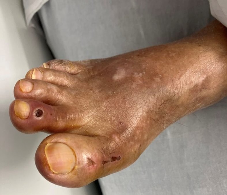
A 77 year-old male presented with new-onset right lower extremity rest pain and ischemic ulcers of the right foot (image 1), consistent with CLTI. His medical history was notable for coronary artery disease status post coronary artery bypass grafting, hypertension, hyperlipidemia, non-insulin-dependent diabetes mellitus with poor glycemic control (hemoglobin A1c of 9.6) and bilateral carotid artery stenosis. He had a long-standing history of PAD and had previously undergone a left below-knee amputation, after which he remained ambulatory with a prosthetic limb. On examination, the patient was well-appearing, alert and oriented. Femoral pulses were palpable bilaterally, but pedal pulses in the right lower extremity were absent. Laboratory studies were unremarkable, with a white blood cell count of 7.0 and a creatinine of 1.18. Noninvasive vascular studies confirmed severe tibial disease.
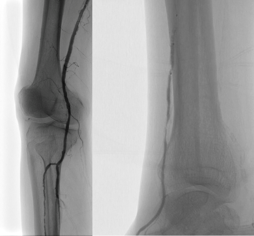
Diagnostic angiography
Diagnostic angiography revealed single-vessel runoff into the foot with an occluded peroneal artery and an incomplete plantar arch from the posterior tibial artery (image 2 & 3). The anterior tibial artery demonstrated severe, heavily calcified disease along its course, representing the primary target for intervention given the limited outflow options.
Interventional procedure
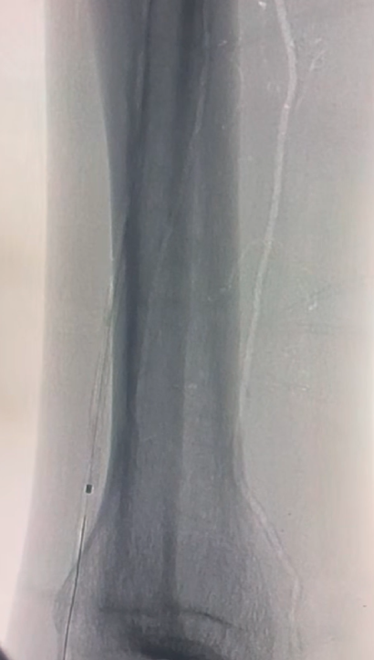
Arterial access was obtained via a contralateral femoral approach using an up-and-over technique with placement of a destination sheath. The lesion was crossed using a 0.014-inch guidewire supported by a crossing catheter. In the proximal anterior tibial artery, a two-millimeter plain old balloon angioplasty was performed, with vessel sizing guided by intravascular ultrasound (IVUS). However, in the distal anterior tibial artery, the operator encountered significant difficulty tracking any treatment balloon through the calcified segment. At this point, the Shockwave Javelin Peripheral IVL catheter was introduced (image 4.) with gentle forward pressure, successfully traversing the lesion and delivering lithotripsy energy to modify the circumferential calcium. Following IVL, post-dilation was completed using a 1.5-millimeter plain old balloon angioplasty catheter. IVUS imaging performed before and after the procedure (image 5. & 6.) confirmed effective calcium modification, with improved luminal gain and vessel compliance in the treated segments.
Outcome
The completion angiogram demonstrated a satisfactory result with restored inline flow to the foot. On follow-up, the patient’s ischemic wounds showed progressive healing, and he remained ambulatory, successfully preserving his right lower extremity and maintaining functional independence. Given that this patient had already undergone a contralateral below-knee amputation, limb salvage of the remaining extremity was of critical importance for his quality of life and mobility.
Discussion
This case highlights several important teaching points for the interventional community. First, severely calcified tibial vessels can render conventional balloon angioplasty ineffective due to the inability to track devices through rigid and occlusive arterial segments. Shockwave Javelin delivered on the novel Forward IVL Platform offers a mechanism to modify intimal and medial calcium beyond the catheter tip, thereby improving vessel compliance, enabling further interventional treatment and achieving the goal of revascularization.
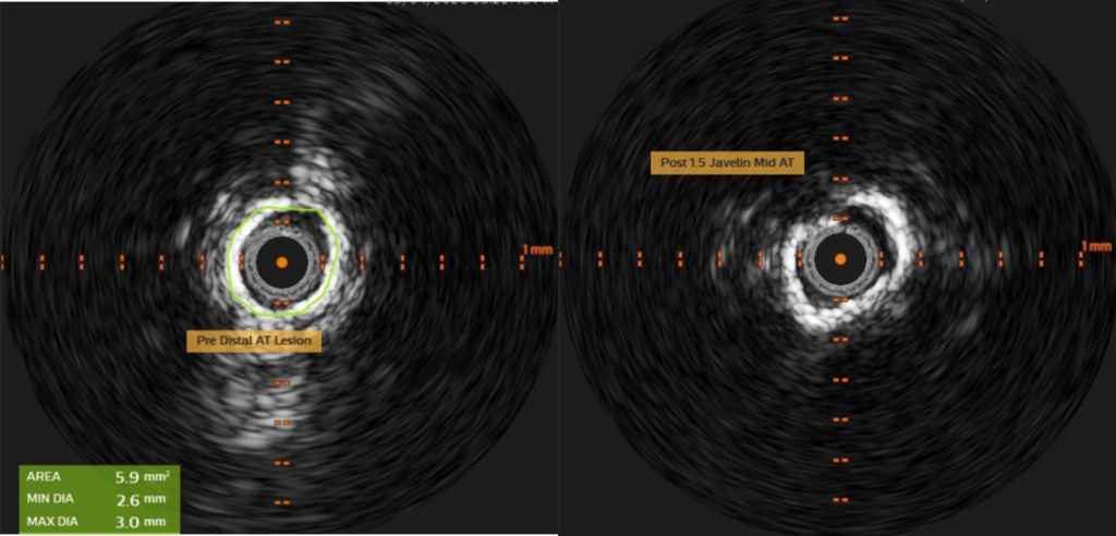
Second, IVUS imaging played a dual role in this case, both for appropriate vessel sizing in the proximal segment and for confirming the effectiveness of calcium modification after lithotripsy. Finally, this case underscores the clinical value of Shockwave Javelin in the tibial territory for patients with CLTI, particularly when the stakes of limb loss are magnified by a contralateral amputation and multiple systemic comorbidities.
Utilization of Shockwave Javelin in severely calcified distal tibial artery disease can lead to safe and effective calcium modification and thus reduce major amputation risks.
Sung Yup (Pablo) Kim, MD, is a board-certified vascular surgeon at The Mount Sinai Hospital and an Associate Professor of Surgery at the Icahn School of Medicine at Mount Sinai. Akila Pai, MD, is a vascular surgeon at The Mount Sinai Hospital. They specialize in the treatment of complex PAD, CLTI, limb salvage and advanced endovascular therapies.
References
1. Corl JD, Clair D, Mwipatayi P, et al. FORWARD PAD IDE/Feasibility Studies: Primary
Endpoint Analysis of a Novel Non-Balloon-Based Peripheral IVL Catheter. JACC:
Cardiovascular Interventions. Published online November 4, 2024. doi:10.1016
jcin.2024.10.035.
2. Corl J, VIVA Late Breaking Clinical Trial 2024.
Sung Yup Kim is a paid consultant of Shockwave Medical. The views expressed are those of the authors and not necessarily those of Shockwave Medical.
Shockwave Javelin Peripheral IVL Catheter Safety Information
In the United States: Rx only.
Indications for Use — The Shockwave Medical IVL System with the Javelin Peripheral IVL Catheter
is intended for lithotripsy- enabled modification and crossing of calcified lesions in the peripheral
vasculature, including the iliac, femoral, ilio-femoral, popliteal, and infra-popliteal arteries, prior to
final treatment.
Not for use in coronary, carotid, or cerebral vasculature. Additionally, not for use in pulmonary vasculature
in the U.S. and New Zealand.
Contraindications — Do not use if unable to pass 0.014″ (0.36 mm) guidewire across the treatment
site-Not intended for treatment of in-stent restenosis or in coronary, carotid, cerebral or pulmonary
arteries.
Warnings — Only to be used by physicians who are familiar with interventional vascular procedures—
Physicians must be trained prior to use of the device— Use the generator in accordance with
recommended settings as stated in the Operator’s Manual.
Precautions — Avoid applying acoustic pressure pulses while IVL window is not filled with sterile saline—
Appropriate anticoagulant therapy should be administered by the physician— Decision regarding use of
distal protection should be made based on physician assessment of treatment lesion morphology.
Adverse effects — Possible adverse effects consistent with standard angioplasty include-Access site
complications -Allergy to contrast or blood thinner- Arterial bypass surgery—Bleeding complications—
Death— Fracture of guidewire or device—Hypertension/Hypotension— Infection/sepsis—Placement
of a stent—renal failure— Shock/pulmonary edema—target vessel stenosis or occlusion— Vascular
complications. Risks unique to the device and its use— Allergy to catheter material(s)— Device
malfunction or failure.
Prior to use, please reference the Instructions for Use for more information on indications,
contraindications, warnings, precautions and adverse events. www.shockwavemedical.com/IFU
© Shockwave Medical, Inc. All Rights Reserved. SPL 83146 Rev. A.